The human side of veterinary medicine
Author: Jeff Budlong
Author: Jeff Budlong

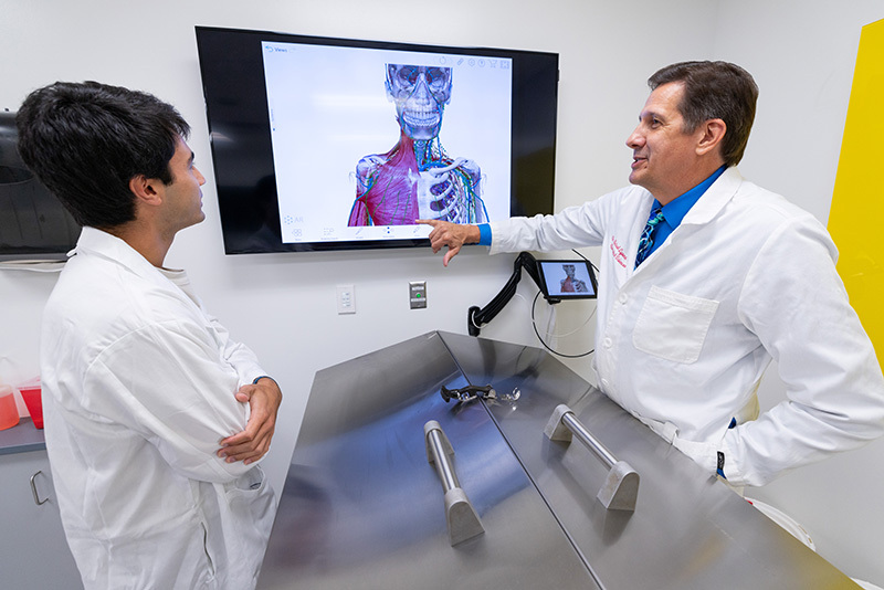
Biomedical sciences teaching professor Michael Lyons (right) uses a a flat-panel screen above a cadaver table during his discussion with a student in the human anatomy lab. Photos by Christopher Gannon.
Innovation can be found all over the College of Veterinary Medicine, even in storage rooms.
To aid students interested in human medicine, biomedical sciences teaching professor Michael Lyons and program specialist Bill Robertson converted a former storage room into a human anatomy lab to elevate the impact of the department's one-year master's degree. The intensive two-semester program, first offered in 2013, prepares students to work in industry, academia or continue their veterinary or human medical school education. Originally, students learned anatomy using dog cadavers, but with many students interested in medical school there was a need for a human anatomy lab.
"When Dr. Lyons was hired, he was adamant: If we're going to prepare students for professional health programs, we need human cadavers," Robertson said.
That became a reality in 2017.
Creating the human anatomy lab required overcoming two challenges: providing sufficient ventilation while maintaining a quiet environment for learning.
"Cadavers are embalmed with a solution that contains formaldehyde, so one of the things you must do is exhaust those fumes," Robertson said. "You could use a huge fan, but it would make so much noise students wouldn't be able to hear themselves think -- that doesn' make for a good learning environment."
To move enough air to eliminate the odor, Robertson and Lyons looked through the literature and found studies that used stainless steel dissection tables equipped with a downdraft exhaust feature. These tables draw fumes through perimeter ventilation slots, funneling them into a ductwork system connected to the building's exhaust.
"To accommodate the ventilation needs, we had walls constructed to provide space for new exhaust ductwork," Robertson said. "We used large ductwork and custom fittings to move a large volume of air at a low velocity while students are working.”
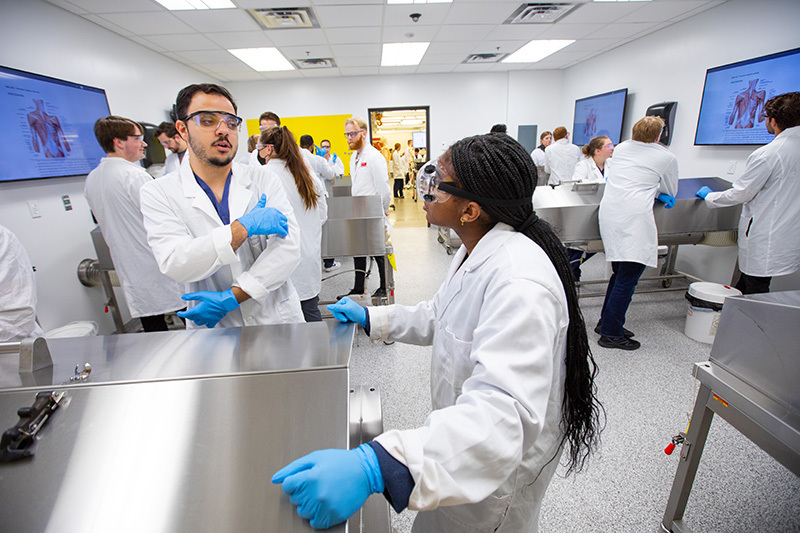
During labs, four students study a cadaver on one of six stainless steel tables. The tables have lids that open to expose the cadaver, and a button on the wall is depressed to increase airflow. Above each table is a wall-mounted flat-panel screen for viewing a dissection and an iPad loaded with a 3D anatomy program.
"When students are not in the lab, it's still necessary to ventilate the tables, but at a lower rate," said Lyons, who noted the delicate balance to avoid drying out the cadavers.
Environmental health and safety staff test the lab -- which remains at about 67 degrees -- twice a year to ensure the air quality meets safety standards. How successful is the design?
"When they measure the air quality in the human lab, formaldehyde fumes are below detection levels because the ventilation is so good," Robertson said. "We succeeded; there is no odor and the room is quiet."
Equally impressive is how fast the space was remodeled and readied for students. Construction began in May 2017 and was completed by fall semester.

The human anatomy lab allows four students to study a cadaver on one of six stainless steel tables. A storage room was transformed into the lab in 2017.
Lyons, who drew from his own experience during his Ph.D. years at the University of Iowa, teaches human anatomy to graduate students in the fall and undergraduates in the spring. Graduate students learn how to dissect during the fall semester, and undergraduates study those dissected donors in the spring.
"It really is about making the most out of the precious gift these donors provide," he said.
The University of Iowa Deeded Body Program provides embalmed human donors to Iowa State each August. At the end of the spring semester, the donors are returned to the University of Iowa, where they are cremated and a memorial service is held. The cremains are then returned to the donor's family. Lyons sets the tone in his class with one unbreakable rule.
"We tell our students these are human beings, and it doesn't matter that they are deceased," he said. "They are people, so treat them with respect. Any family member of someone who has been donated expects that, and we do, too."
Human anatomy classes are in such demand the available spots fill up quickly each semester. The master's program in athletic training added the human anatomy class to its curriculum, so now the class is offered during the first half of summer and also is open to additional undergraduate and graduate students. The demand for health care professionals demonstrates the need for additional anatomy classes.
Because of space restrictions, during lab time, half of the students first work in the human lab and the other half in the gross anatomy room where veterinary students learn animal anatomy. Then the two groups switch.
Graduate students spend two hours in lecture and 7.5 hours in the lab weekly, while undergrads have two hours of lecture and four hours of lab work. Many students spend extra time in the lab but are required to work with a partner. A ceiling camera continuously monitors the lab, and only those with key cards have access. When they're done, students wipe down tables, mop floors and apply wetting solution to the bodies.
Lyons was honored with a university award for his teaching in 2024. In their nomination letters, students who went on to medical school praised Lyons' instruction for preparing them to excel.
"I focus on skills and how to think like an anatomist because that is where most students end up having the biggest issues, based on feedback from medical school anatomists," Lyons said. "The class is designed to help students deal with the amount of information they encounter, but at the same time, synthesize the material to make clinical applications."
He incorporates real-life examples so students must apply information they are learning and avoid relying on memorization. He also emphasizes learning how each organ, muscle or vein interacts with what's around it. Graduate students also get dissection experience, a skill not taught in a professional setting, Lyons said.
Lyons is the lone faculty instructor for the courses, but he uses recent graduates as lab instructors and grad students who excel during the fall work as teaching assistants in the spring.
"It allows them to keep working on the anatomy and get more experience," he said. "Many go on to be teaching assistants in their professional program."
The only restriction on growing the human anatomy program is space. Adjacent rooms aren't available for expansion, but that doesn't stop Lyons from imagining what a larger program could mean for students and the university.
"Not everyone understands the relationship between human and animal health," he said. "It's the concept of One Health, a more holistic look at health, which I think is a huge advantage to Iowa State."
Lyons thinks Colorado State University may be the only other veterinary medicine college with a human anatomy lab. He hopes to partner with veterinary faculty to create more opportunities for future veterinarians and doctors to interact and learn about the links between the two.
"Right now, we offer an optional experience at the end of first-year vet students' anatomy classes to observe the human donors," Lyons said. "It's neat for them to see something they haven't dissected as part of their training. My students benefit because they can talk to the DVM students about similarities and differences in human and animal anatomy. Since the human donors are older, vet students get to see more age-related pathologies than they can see during their first year of anatomy training."